Home
/ Foot Tendon Diagram : Diagram The Ankle - A tendon tear can be painful and make it hard to do any activities that require you to put weight on your foot.
Foot Tendon Diagram : Diagram The Ankle - A tendon tear can be painful and make it hard to do any activities that require you to put weight on your foot.
Foot Tendon Diagram : Diagram The Ankle - A tendon tear can be painful and make it hard to do any activities that require you to put weight on your foot.. The achilles tendon connects the heel to the calf muscle and is essential for running jumping and standing on the toes. The lisfranc joint complex is a series of ligaments that stabilize the tarsometatarsal joints. Fpe medical review board a foot pain diagram is a great tool to help you work out what is causing your ankle and foot pain. Due to less blood flow in ligaments, sprains are not easily recovered and long term damage results on the ligaments. The thick bands of tissues that connect muscles to bones are called tendons.
Overuse, like the repetitive movements of running or playing sports, can cause the posterior tibial tendon to become strained or ruptured. These tendons help your extensor muscles pull your foot upwards, which is necessary for walking. This movement allows us to stand on our toes when walking. A tendon is band of tissue made up of many fibers. There may be trauma associated with a torn tendon of the foot.
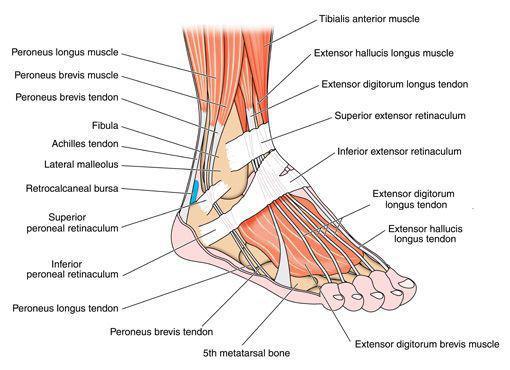
Foot Anatomy Bones Ligaments Muscles Tendons Arches And Skin from biologydictionary.net Hand tendons diagram, picture of hand tendons diagram. Attaches the calf muscles to the calcaneus, most important muscles for running, jumping, walking etc. The achilles tendon is the largest and strongest tendon in the body. Hochwertige kletterseile für dein outdoor abenteuer! Tendon diagram of calf and knee. Problems such as flat feet or high arches can create muscular. One peroneal tendon attaches to the outer part of the midfoot, while the other tendon runs under the foot and attaches near the inside of the arch. Ligaments sprains are more common, but severe injuries.
The human foot combines mechanical complexity and structural strength.
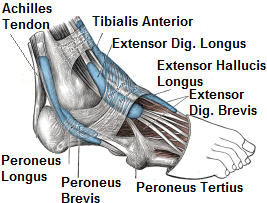
Foot tendon anatomy diagram get rid of wiring diagram problem. The foot has a number of tendons. Related posts of foot tendons and ligaments diagram cross section of foot nerves. Allows the foot to be turned inward and also supports the arch of the foot. The peroneal tendons run down together behind the outer side of the ankle and then split before attaching to different parts of the foot. The muscles are located mainly in the sole of the foot and divided into a central (medial) group and a group on either side (lateral). The plantar ligaments are stronger than those on the dorsal side (figure 12 & 13). A tendon is band of tissue made up of many fibers. Other tendons help to control the movements of the toes. Allows the action of raising the foot. Attaches the calf muscles to the calcaneus, most important muscles for running, jumping, walking etc. Many tendons attach these muscles to the bones and ligaments that hold the bones together to maintain the foot's arch. The calcaneus (heel bone) is the largest bone in the foot.
Tendon diagram of calf and knee. The calcaneus (heel bone) is the largest bone in the foot. The tendons are thick bands that connect muscles to bones. A tendon is a band of tissue that connects a muscle to a bone. Foot tendon anatomy diagram get rid of wiring diagram problem.
Foot And Ankle Sportsmed from sportsmedalabama.com When the calf muscles flex, the achilles tendon pulls on the heel. The plantar ligaments are stronger than those on the dorsal side (figure 12 & 13). Overuse, like the repetitive movements of running or playing sports, can cause the posterior tibial tendon to become strained or ruptured. Problems such as flat feet or high arches can create muscular. Other tendons help to control the movements of the toes. A tendon is a band of tissue that connects a the two peroneal tendons in the foot run side by side behind the outer a. These ligaments were described by the napoleonic. Ligaments sprains are more common, but severe injuries.
One of the main ligaments in the foot is the plantar fascia, which forms the arch on the sole of the foot.
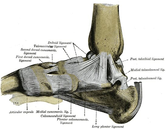
They are remarkably strong, having one of the. Foot tendons and ligaments diagram. The muscles, tendons and ligaments. This diagram shows the medial aspect of the foot. The lisfranc joint complex is a series of ligaments that stabilize the tarsometatarsal joints. They are stronger across the plantar (sole) of the foot than on the dorsal (top) aspect, though they are very strong in either case. Hochwertige kletterseile für dein outdoor abenteuer! Overuse, like the repetitive movements of running or playing sports, can cause the posterior tibial tendon to become strained or ruptured. This diagram shows the sole of the foot. The foot diagram has a complex structure made up of bones, ligaments, muscles, and tendons.understanding the structure of the foot is best done by looking at a foot diagram where the anatomy has been labeled. Hi, for the last week or so, i get this sudden sharp and pulsating pain in my foot, the inner arch area (left feet).i looked at a foot muscle diagram to identify the area and it looks like the abductor hallucis area where the pain is located. Foot tendon anatomy diagram get rid of wiring diagram problem. Allows the action of raising the foot.
It connects muscle to bone. The foot has a number of tendons. A foot tendon tear happens when one of the tendons in the foot is damaged from sudden injury or overuse. Tendon tissue is also known as sinew. This may be caused by sudden trauma, such as rolling the foot, which causes the ligaments to pull away from the bone.
Foot Ankle Tendonitis Causes Symptoms Treatment from www.foot-pain-explored.com Originates from the lower part of the fibula and attaches to the outer side of the midfoot Runners are often subject to this painful condition. The calcaneus (heel bone) is the largest bone in the foot. The muscles at the top of the foot fan out to supply the individual toes. Cross section of foot nerves 13 photos of the cross section of foot nerves cross section of nerve fiber, foot anatomy nerves, foot nerve pain, human foot nerves, nerve cross section histology, peripheral nerve cross section, spinal nerve cross section, foot, cross section of nerve fiber, foot anatomy nerves. Muscles of the foot dorsal plantar. Due to less blood flow in ligaments, sprains are not easily recovered and long term damage results on the ligaments. Also allows the action of raising up onto toes.
Originates from the lower part of the fibula and attaches to the outer side of the midfoot
Foot anatomy bones ligaments muscles tendons arches. The lisfranc joint complex is a series of ligaments that stabilize the tarsometatarsal joints. A tendon is a band of tissue that connects a muscle to a bone. The foot diagram has a complex structure made up of bones, ligaments, muscles, and tendons.understanding the structure of the foot is best done by looking at a foot diagram where the anatomy has been labeled. Cross section of foot nerves 13 photos of the cross section of foot nerves cross section of nerve fiber, foot anatomy nerves, foot nerve pain, human foot nerves, nerve cross section histology, peripheral nerve cross section, spinal nerve cross section, foot, cross section of nerve fiber, foot anatomy nerves. The achilles tendon connects the heel to the calf muscle and is essential for running jumping and standing on the toes. Hi, for the last week or so, i get this sudden sharp and pulsating pain in my foot, the inner arch area (left feet).i looked at a foot muscle diagram to identify the area and it looks like the abductor hallucis area where the pain is located. Foot tendon anatomy diagram get rid of wiring diagram problem. The tendons are thick bands that connect muscles to bones. Overuse, like the repetitive movements of running or playing sports, can cause the posterior tibial tendon to become strained or ruptured. Bones, muscles, tendons and nerves which will each give slightly different foot pain symptoms. Muscles, tendons, and ligaments run along the surfaces of the feet, allowing the complex movements needed for motion and balance. Chloe wilson bsc(hons) physiotherapy reviewed by:
Bones, muscles, tendons and nerves which will each give slightly different foot pain symptoms tendon diagram. Ligaments hold bones together and stabilize joints.